Microcystis aeruginosa
blue green algae
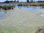
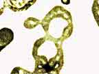
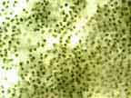
Microcystis aeruginosa is a unicellular or colonial cyanobacteria. It produces a blue pigment that enables it to be clearly seen in the event of an algal bloom. The algae may multiply to such an extent in the summer months as to discolour the water so it appears green, blue green, or greenish brown. The algae may release toxins into the water.
Other blue green algae.
Worldwide in natural or induced eutrophic (nutrient enriched/ stagnant) water bodies. Also found in still fresh waters, such as lakes, ponds, canals and reservoirs.
Cyanotoxins include cyclic peptides, alkaloids and lipopolysaccharides (LPS). Hepatotoxic cyclic peptides (eg microcycystin and nodularin) are found most frequently. The alkaloids (eg anatoxin a, saxitoxins) tend to be neurotoxic and very little work has been done with the LPS toxin. Toxins are produced by the algae and released into the water when they die.
Microcystin causes the breakdown of the sinusoidal endothelium, damage to hepatocyte membranes, and necrotic changes in hepatocyte cytoplasm. Death is considered to be related to shock following massive liver necrosis and haemorrhage.
All mammals. Sickness and death has been reported in livestock, pets birds, and wildlife following ingestion.
Toxic signs may appear within 1hr, and death may occur in less than 24hrs after ingestion of the poisonous material. The most commonly reported sequence of events is rapid prostration, convulsions, and death. Microcystin may cause abdominal pain, anorexia, vomiting, muscular tremors, dyspnea, cyanosis and salivation are common; less common are icterus, diarrhoea, and bloody faeces. Photosensitisation frequently occurs in animals that survive for several days.
Microcystis aeruginosa toxicity results in a markedly enlarged and often haemorrhagic liver, fluid accumulation in the body cavities, and widespread petechiae and ecchymoses. In peracute cases the liver is swollen, friable and congested. In acute cases the periacinar hepatocytes are necrotic with haemorrhage, while the remainder of the acinus shows hydropic degeneration or fatty change. Oedema and haemorrhage of the gall bladder wall may also be prominent. Nephrosis, splenomegaly, congestion of the viscera, lung oedema and fatty change in the myocardium are other lesions which may be encountered Microscopic examination reveals centrilobular to near massive necrosis. In chronic cases, hepatic cirrhosis, icterus, photosensitivity, emaciation and ascites may be present.
Diagnosis is based on history (recent contact with an abundance of potentially toxic freshwater algae), clinical signs and postmortem findings. Samples of the algae should be collected as soon as possible to confirm the toxicity of the strain in the laboratory. Marked rises in serum concentration of liver enzymes and bilirubin, and changes in coagulation parameters have been reported.
Other toxicities causing liver failure or neurological signs.
Removal from the source, place animals in a protected area away from direct sunlight. Provide ample water and good quality feed. Activated charcoal and a laxative dose of mineral oil (not given concurrently with activated charcoal) can help remove the toxin from the GI tract. In surviving animals, antibiotics, glucose, calcium and magnesium supplementation have been used.
Prognosis poor, usually fatal or severe illness. A long recuperation period can be expected for animals that survive.
Check water supplies regularly in the summer.
Wood, S. 2001. Cyanobacteria an underestimated risk to animal health in New Zealand? VetScript. December XIV 11:4 5.
Surveillance, Blue-green algae toxicosis, 35 (2) 32 2008
Surveillance, Cyanobacterial toxicity, 35 (4) 13 2008
4 October, 2007